

Mitochondrial dysfunction induced by PM2.5 in human umbilical vein endothelial cells
-
摘要:
目的 探讨颗粒物空气动力学直径≤ 2.5 μm(particulate matter 2.5,PM2.5)对人脐静脉内皮细胞EA.hy926细胞线粒体功能损伤机制。 方法 用交通相关PM2.5(0、25、50、100 μg/mL)处理EA.hy926细胞24 h,观察其对EA.hy926细胞线粒体功能的损伤。 结果 PM2.5处理24 h后,EA.hy926细胞表现出线粒体三磷酸腺苷(adenosine triphosphate,ATP)产量和线粒体DNA(mitochondrial DNA,mtDNA)水平下降,线粒体结构完整性破坏等功能紊乱。PM2.5处理也可诱导增加活性氧(reactive oxygen species,ROS)产生,线粒体膜电位(mitochondrial membrane potential,ΔΨm)下降。此外,用PM2.5处理EA.hy926细胞可导致过氧化物酶体增殖物激活受体γ辅激活子1α(peroxisome proliferator-activated receptor gamma coactivator 1-alpha,PGC-1α)的mRNA表达减少、线粒体转录因子A(mitochondrial transcription factor A,TFAM)的mRNA表达和parkin与动力蛋白1样蛋白(parkin and dynamin 1-like protein 1,Drp1)蛋白表达增加,但PGC-1α蛋白表达降低。 结论 在EA.hy926细胞中,PM2.5诱导线粒体功能损伤作用可能是通过促进ROS产生,以及诱导自噬和线粒体生物发生功能障碍介导的。 -
关键词:
- PM2.5 /
- EA.hy926细胞 /
- 线粒体功能损伤
Abstract:Objective To investigate the mechanism of mitochondrial dysfunction of human umbilical vein endothelial cell EA. Hy926 cells. Methods EA. Hy926 cells were treated with traffic-related particulate matter 2.5(PM2.5)(0, 25, 50, 100 μg/mL) for 24 h, the damage of PM2.5 on mitochondrial function. Results EA. Hy926 cells treated with PM2.5 for 24h, decreased mitochondrial ATP production and mitochondrial DNA(mtDNA) level, and destruction of mitochondrial structural integrity. PM2.5 treatment also induced the increase in reactive oxygen species (ROS) generation, decline in mitochondrial membrane potential (ΔΨm).In addition, the treatment of EA. Hy926 cells with PM2.5 resulted in decreased mRNA expression of Peroxisome proliferator-activated receptor gamma coactivator 1-alpha (PGC-1α), increased mRNA expression of mitochondrial transcription factor A(Tfam), and increased the protein expression of parkin and dynamin 1-like protein 1 (Drp1) in the EA. Hy926 cells, but decreased the protein expression of PGC-1α. Conclusions PM2.5 can induce mitochondrial dysfunction in EA. Hy926 cells and these effects are mediated through the promotion of ROS overproduction, as well as the promotion of mitochondrial autophagy and mitochondrial biogenesis dysfunction. -
Key words:
- PM2.5 /
- EA.hy926 /
- Mitochondrial dysfunction
-

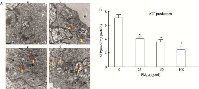
图 1 PM2.5对EA.hy926细胞线粒体形态和ATP产生功能的损伤效应
注:图 1. 用PM2.5(25, 50, 100 μg/mL)处理EA.hy926细胞24 h表现出线粒体功能障碍。A.a)未处理的EA.hy926细胞; 线粒体形态保持完整;b) PM2.5(25 μg /mL)处理EA.hy926细胞24 h;c) PM2.5(50 μg /mL)处理EA.hy926细胞24 h; d)PM2.5(100 μg /mL)处理EA.hy926细胞24 h。箭头指示线粒体,S,肿胀,V空泡形成,C,线粒体嵴消失。比例尺, 0.5 μm. B. PM2.5对细胞ATP产生的影响。3个独立实验,数据表示x±s。与对照组相比,*P<0.05,与PM2.5(50 μg /mL)处理组相比,#P<0.05。
Figure 1. The mitochondrial morphology and ATP production function of EA. Hy926 cells damaged by PM2.5

图 2 PM2.5诱导过度产生ROS和降低ΔΨm
注:(A)用共聚焦显微镜成像检测线粒体膜电位ΔΨm (原始放大,400×)。橙色表示绿色(JC-1单体)与红色(JC-1集合体)的结合。(a)对照组,未处理的EA.hy926细胞; (b) PM2.5(2 5μg /mL) 处理EA.hy926细胞24 h; (c) PM2.5(50 μg /mL) 处理EA.hy926细胞24 h; (d) PM2.5(100 μg /mL) 处理EA.hy926细胞24 h。(B)定量分析PM2.5对ROS产生和(C)线粒体膜电位ΔΨm作用。数据用(x±s)表示,3次独立实验。 & 与PM2.5(25 μg /mL)处理组相比,P<0.05;#与PM2.5(50 μg /mL)处理组相比,P<0.05。
Figure 2. PM2.5 induced overproduction of ROS and decreased ΔΨm

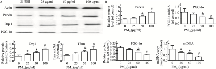
图 3 PM2.5对EA.hy926细胞线粒体自噬和生物发生相关基因表达及mtDNA拷贝数的影响
注:A.parkin, Drp1和PGC-1α的蛋白表达。B.(a)Parkin蛋白表达,(b)Drp1蛋白表达,(c)PGC-1α蛋白表达,(d) PGC-1αmRNA表达,(e).Tfam mRNA表达和f.mtDNA拷贝数。数据用(x±s)表示,3次独立实验。与对照组相比,*P<0.05,与PM2.5(25 μg /mL)处理组相比, & P<0.05;与PM2.5(50 μg /mL)处理组相比,#P<0.05。
Figure 3. Effects of PM2.5 on mitochondrial autophagy, mitochondrial biogenesis related gene expression and mtDNA copy number in EA. Hy926 cells
表 1 PM2.5诱导EA.hy926细胞线粒体功能异常相关指标相对表达量(x±s)
Table 1. Relative expression levels of related indicators of mitochondrial dysfunction in EA. Hy926 cells induced by PM2.5(x±s)
相关指标 对照组a 25 μg/ml处理组b 50 μg/ml处理组c 100 μg/ml处理组d F值 P值 ATP 7.100±0.400 4.067±0.231 3.567±0.321 2.467±0.513 81.33 < 0.001 ROS 0.034±0.018 0.051±0.021 0.068±0.021 0.117±0.036 6.12 0.018 ΔΨm 0.519±0.148 0.294±0.145 0.193± 0.119 0.059±0.040 7.72 0.010 mtDNA 1.000±0.000 0.709±0.098 0.620±0.059 0.321±0.060 56.18 < 0.001 Drp1蛋白 0.222±0.056 0.303±0.062 0.394±0.051 0.539±0.076 14.38 0.001 Parkin蛋白 0.194±0.059 0.272±0.031 0.369±0.021 0.548±0.067 29.9 < 0.001 PGC-1α蛋白 0.191±0.020 0.163±0.013 0.123±0.020 0.084±0.010 24.59 < 0.001 PGC-1α mRNA 1.039±0.326 0.846±0.135 0.616±0.087 0.437±0.012 6.29 0.017 Tfam mRNA 1.009±0.161 1.263±0.293 1.662±0.204 2.064±0.539 5.78 0.021 注:a未处理的EA.hy926细胞; b用25 μg/ml PM2.5处理EA.hy926细胞24 h; c用50 μg/ml PM2.5处理EA.hy926细胞24 h; d用100 μg/ml PM2.5处理EA.hy926细胞24 h。  下载: 导出CSV
-
[1] 中华人民共和国卫生部. 中国卫生统计年鉴2009-2012. [M]. 北京: 中国协和医科大学出版社, 2016.Ministry of Health of the People's Republic of China. China health statistics yearbook 2009-2012. [M]. Beijing: China Union Medical College Press, 2016. [2] 陈伟伟, 高润霖, 刘力生, 等. 《中国心血管病报告2017》概要[J]. 中国循环杂志, 2018, 33(1): 1-8. DOI: 10.3969/j.issn.1000-3614.2018.01.001.Chen WW, Gao RL, Liu LS, et al. Summary of "China cardiovascular disease report 2017"[J]. Chinese Circulation Journal, 2018, 33(1): 1-8. DOI: 10.3969/j.issn.1000-3614.2018.01.001. [3] 孙娜, 刘启玲, 张荣强, 等. 大气污染物对成人和儿童外周血基因表达谱的影响及生物信息学分析[J]. 中华疾病控制杂志, 2018, 22(7): 677-681. DOI: 10.16462/j.cnki.zhjbkz.2018.07.006Sun N, Liu QL, Zhang RQ, et al. Genome-wide differential gene expression in children and adults exposed to air pollution[J]. Chin J Dis Control Prev, 2018, 22(7): 677-681. DOI: 10.16462/j.cnki.zhjbkz.2018.07.006 [4] Yu E, Calvert PA, Mercer JR, et al. Mitochondrial DNA damage can promote atherosclerosis independently of reactive oxygen species through effects on smooth muscle cells and monocytes and correlates with higher-risk plaques in humans[J]. Circulation, 2013, 128(7): 702-712. DOI: 10.1161/CIRCULATIONAHA.113.002271. [5] Mercer JR. Mitochondrial bioenergetics and therapeutic intervention in cardiovascular disease[J]. Pharmacol Ther, 2014, 141(1): 13-20. DOI: 10.1016/j.pharmthera.2013.07.011. [6] Ribeiro JP, Kalb AC, Campos PP, , et al. Toxicological effects of particulate matter (PM2.5) on rats: Bioaccumulation, antioxidant alterations, lipid damage, and ABC transporter activity[J]. Chemosphere, 2016, 163: 569-577. DOI: 10.1016/j.chemosphere.2016.07.094. [7] Deng X, Rui W, Zhang F, et al. PM2.5 induces Nrf2-mediated defense mechanisms against oxidative stress by activating PIK3/AKT signaling pathway in human lung alveolar epithelial A549 cells[J]. Cell Biol Toxicol, 2013, 29(3): 143-157. DOI: 10.1007/s10565-013-9242-5. [8] Deng X, Zhang F, Rui W, et al. PM2.5-induced oxidative stress triggers autophagy in human lung epithelial A549 cells[J]. Toxicol In Vitro, 2013, 27(6): 1762-1770. DOI: 10.1016/j.tiv.2013.05.004. [9] Cimellaro A, Perticone M, Fiorentino TV, et al. Role of endoplasmic reticulum stress in endothelial dysfunction[J]. Nutr Metab Cardiovasc Dis, 2016, 26(10): 863-871. DOI: 10.1016/j.numecd.2016.05.008. [10] Yang GZ, Wang ZJ, Bai F, et al. Epigallocatechin-3-gallate protects HUVECs from PM2.5-induced oxidative stress injury by activating critical antioxidant pathways[J]. Molecules, 2015, 20(4): 6626-6639. DOI: 10.3390/molecules20046626. [11] Cui Y, Xie X, Jia F, et al. Ambient fine particulate matter induces apoptosis of endothelial progenitor cells through reactive oxygen species formation[J]. Cell Physiol Biochem, 2015, 35(1): 353-363. DOI: 10.1159/000369701. [12] Li R, Kou X, Geng H, et al. Effect of ambient PM(2.5) on lung mitochondrial damage and fusion/fission gene expression in rats[J]. Chem Res Toxicol, 2015, 28(3): 408-418. DOI: 10.1021/tx5003723. [13] Xu Z, Xu X, Zhong M, et al. Ambient particulate air pollution induces oxidative stress and alterations of mitochondria and gene expression in brown and white adipose tissues[J]. Part Fibre Toxicol, 2011, 8: 20. DOI: 10.1186/1743-8977-8-20. [14] Lu Y, Li S, Wu H, et al. Beneficial effects of astragaloside Ⅳ against angiotensin Ⅱ-induced mitochondrial dysfunction in rat vascular smooth muscle cells[J]. Int J Mol Med, 2015, 36(5): 1223-1232. DOI: 10.3892/ijmm.2015.2345. [15] Kageyama Y, Hoshijima M, Seo K, et al. Parkin-independent mitophagy requires Drp1 and maintains the integrity of mammalian heart and brain[J]. EMBO J, 2014, 33(23): 2798-2813. DOI: 10.15252/embj.201488658. [16] Wu Z, Puigserver P, Andersson U, et al. Mechanisms controlling mitochondrial biogenesis and respiration through the thermogenic coactivator PGC-1[J]. Cell, 1999, 98(1): 115-124. DOI: 10.1016/S0092-8674(00)80611-X. [17] Xie H, Lev D, Gong Y, et al. Reduced mitochondrial DNA copy number in peripheral blood leukocytes increases the risk of soft tissue sarcoma[J]. Carcinogenesis, 2013, 34(5): 1039-1043. DOI: 10.1093/carcin/bgt023. [18] Narendra DP, Youle RJ. Youle Targeting mitochondrial dysfunction: role for PINK1 and Parkin in mitochondrial quality control[J]. Antioxid Redox Signal, 2011, 14(10): 1929-1938. DOI: 10.1089/ars.2010.3799. [19] Deng H, Dodson MW, Huang H, et al. The Parkinson's disease genes pink1 and parkin promote mitochondrial fission and/or inhibit fusion in Drosophila[J]. Proc Natl Acad Sci USA, 2008, 105(38): 14503-14508. DOI: 10.1073/pnas.0803998105. [20] Scarpulla RC, Vega RB, Kelly DP. Transcriptional integration of mitochondrial biogenesis[J]. Trends Endocrinol Metab, 2012, 23(9): 459-466. DOI: 10.1016/j.tem.2012.06.006. -


 点击查看大图
计量
- 文章访问数: 320
- HTML全文浏览量: 127
- PDF下载量: 20
- 被引次数: 0