Evaluation of the value of immature reticulocyte fraction in the diagnosis of viral diseases in children
-
摘要:
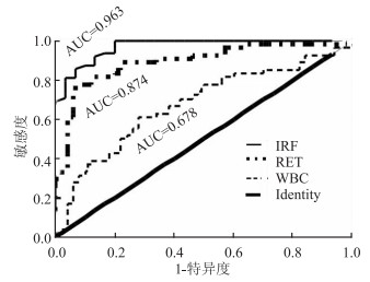
目的 本文主要研究未成熟网织红细胞比率(immature reticulocyte fraction,IRF)测定在儿童病毒性疾病诊断中的价值。 方法 应用流式细胞术对已证实的儿科病毒性感染与细菌性感染各48名急性患儿及40例正常对照组儿童的IRF进行测定,同时对网织红细胞(reticulocyte,RET)、IRF、白细胞(white blood cell,WBC)等进行受试者操作特征曲线(receiver operating characteristic curve,ROC)分析。 结果 病毒感染组IRF(2.05±1.20)明显低于细菌感染组(8.17±2.98)(F=2.89,P=0.007)及正常对照组(8.52±3.57)(F=2.84,P=0.008)。用ROC曲线分析,IRF的曲线下面积(area under curve,AUC)为0.963,RET的AUC为0.874,WBC的AUC为0.678。IRF的最佳临界值为2.45,其敏感度为91.7%,特异度为87.5%,诊断正确率为89.6%。 结论 病毒感染患儿是以IRF下降为特征的;IRF结合WBC等指标可预估儿童早期感染是病毒感染还是细菌感染。 -
关键词:
- 未成熟网织红细胞比率 /
- 感染性疾病 /
- 细菌感染 /
- 病毒感染 /
- ROC曲线
-
表 1 各组5项指标结果比较
Table 1. Comparison of results of 5 indicators in each group
组别 n 白细胞计数(×109/L) 中性粒细胞分类(%) 淋巴细胞分类(%) IRF(%) RET(%) 病毒感染组 48 6.36±2.06a 52.62±18.97a 39.59±17.68a 2.05±1.20ab 0.62±0.43ab 细菌感染组 48 12.49±4.03b 68.14±16.78b 24.85±14.25b 8.17±2.98 1.02±0.36 正常对照组 40 7.60±2.10 58.61±7.25 33.32±6.18 8.52±3.57 1.07±0.36 F值 58.076 37.581 65.897 83.057 18.074 P值 < 0.001 < 0.001 < 0.001 < 0.001 < 0.001 注:a与细菌组比较P < 0.05;b与对照组比较P < 0.05。 -
[1] Morkis IV, Farias MG, Scotti L. Determination of reference ranges for immature platelet and reticulocyte fractions and reticulocyte hemoglobin equivalent[J]. Rev Bras Hematol Hemoter, 2016, 38(4): 310-313. DOI: 10.1016/j.bjhh.2016.07.001. [2] Nadarajan VS, Ooi CH, Sthaneshwar P, et al. The utility of immature reticulocyte fraction as an indicator of erythropoietic response to altitude training in elite cyclists[J]. Int J Lab Hematol, 2010, 32(1 pt2): 82-87. DOI:10. 1111/j.1751-553X.2008.01132.x. [3] 周凤玲, 靳桂明, 董玉梅, 等. 儿童感染性疾病病原菌分布及耐药性分析[J]. 中华医院感染学杂志, 2014, 24(20): 5139-5141. DOI:10. 11816/cn.ni.2014-142814.Zhou FL, Jin GM, Dong YM, et al. Distribution and drug resistance of pathogens causing infectious diseases in children[J]. Chin J Nosocomiol, 2014, 24(20): 5139-5141. DOI:10. 11816/cn.ni.2014-142814. [4] 郝坤艳, 于乐成, 高蕾, 等. 戊型肝炎相关性纯红细胞再生障碍性贫血1例报告[J]. 临床肝胆杂志. 2015, 31(7): 1126-1127. DOI: 10.3969/j.issn.1001-5256.2015.07.031.Hao KY, Yu LC, Gao L, et al. Hepatitis e-associated pure red cell aplasia: a report of one case[J]. J Clin Hepatol, 2015, 31(7): 1126-1127. DOI: 10.3969/j.issn.1001-5256.2015.07.031 [5] 任庆旗, 鞠卫强, 王东平, 等. 肝移植后人细小病毒B19感染导致单纯红细胞再生障碍性贫血一例[J]. 中华器官移植杂志, 2016, 37(3): 144-149. DOI:10. 3760/cmaj.issn. 0254-1785.2016.03.004.Ren QQ, Ju WQ, Wang DP, et al. Diagnosis and treatment of pure red cell aplasia caused by human parvovirus B19 afteralivers transplantation[J]. Chin J Organ Transplant, 2016, 37(3): 144-149. DOI:10. 3760/cmaj.issn. 0254-1785.2016.03.004. [6] 张文雍, 谢利霞, 蒋欢欢, 等. 纯红细胞再生障碍性贫血患者巨细胞病毒感染1例[J]. 中国感染与化疗杂志, 2016, 16(4): 502-503. DOI:10. 16718/j.1009-7708.2016.04.022.Zhang WY, Xie LX, Jiang HH, et al. A case report of cytomegalovirus infection in a patient with pure red cell aplasia[J]. Chin J Infect Chemother, 2016, 16(4): 502-503. DOI:10. 16718/j.1009-7708.2016.04.022. [7] 宋花玲, 贺佳, 黄品贤, 等. ROC曲线下面积估计的参数法与非参数法的应用研究[J]. 第二军医大学学报, 2006, 27(7): 726-728. DOI:10. 16781/j.0258-879x.2006.07.011.Song HL, He J, Huang PX, et al. Application of parametric method and non-parametric method in estimation of area under ROC curve[J]. Acad J Sec Mil Med Univ, 2006, 27(7): 726-728. DOI:10. 16781/j.0258-879x.2006.07.011. [8] 徐林发, 汪素珍, 王柏省. 应用ROC曲线求解最佳切点的方法介绍[J]. 中国卫生统计, 2011, 28(6): 701-702. DOI: 10.3969/j.issn.1002-3674.2011.06.030.Xu LF, Wang SZ, Wang BX. Introduction to the method of solving the best cut point by using ROC curve[J]. Chin J Health Statistics, 2011, 28(6): 701-702. DOI:10. 3969/j.issn.1002-3674.2011.06.030. [9] 马科, 左路广, 冯博, 等. 成人外周血未成熟粒细胞正常参考值的建立与临床应用[J]. 广东医学, 2017, 38(21): 3280-3282. DOI:10. 13820/j.cnki.gdyx. 2017.21.008.Ma K, Zuo LG, Feng B, et al. Establishment and clinical application of normal reference value of adult peripheral blood immature granulocytes[J]. Guangdong Med J, 2017, 38(21): 3280-3282. DOI:10. 13820/j.cnki.gdyx. 2017.21.008. -


 下载:
下载:

