

Consistency study of two methods in detection of human epidermal growth factor receptor-2 in breast cancer
-
摘要:
目的 对比分析免疫组织化学法(immunohistochemistry,IHC)和荧光原位杂交法(fluorescence in situ hybridization,FISH)对乳腺癌人类表皮生长因子受体2(human epidermal growth factor receptor-2,HER-2)检测的一致性评价。 方法 选取2013―2018年入住安徽省某三级甲等医院并确诊患乳腺癌的患者组织,分别采用IHC及FISH两种方法检测HER-2,对比其结果的差异及联系。 结果 在选取的276例乳腺癌患者体内发现FISH法检测HER-2扩增与乳腺癌组织学分级相关。IHC法检测HER-2结果为3+的病例与同时做FISH检测结果为阳性扩增的符合率可以达到90%以上。当IHC为-/+和3+时,2种方法检测结果的一致性最好。而HER-2检测为2+的病人,与FISH检测方法相比,两者的符合率仅为67.9%。 结论 HER-2基因扩增与乳腺癌的恶性程度增高有关,IHC筛查HER-2检测结果为2+乳腺癌需加做FISH检测,对于乳腺癌患者的进一步诊疗具有指导意义。 -
关键词:
- 乳腺癌 /
- 人类表皮生长因子受体2 /
- 免疫组织化学法 /
- 荧光原位杂交法
Abstract:Objective To evaluate the consistency of immunohistochemistry (IHC) and fluorescence in situ hybridization (FISH) for the detection of human epidermal growth factor receptor-2 (HER-2) in breast cancer. Methods The patients who were diagnosed with breast cancer in a third-class A hospital in Anhui Province from 2013 to 2018 were selected. IHC and FISH were used to detect HER-2, and the differences and connections were compared. Results In the selected 276 breast cancer patients, FISH detection of HER-2 amplification was related to the histological grade of breast cancer. For patients with HER-2 of 3+ detected by IHC, the positive amplification rate of FISH could reach higher than 90%. When IHC was -/+ and 3+, the consistency of detection results between the two methods was the best.For patients with 2+ in HER-2 test, compared with FISH test, the coincidence rate of IHC was only 67.9%. Conclusions HER-2 gene amplification is associated with increased malignancy of breast cancer, HER-2 screening results of 2+ breast cancer by IHC requires additional FISH test, which is of guiding significance for the further diagnosis and treatment of patients. -

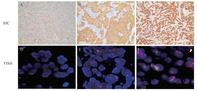
图 1 HER-2蛋白表达代表性染色和FISH检测HER-2基因扩增结果
Figure 1. The results of the representative staining of HER-2 protein expression and HER-2 gene amplification detected by FISH corn
表 1 IHC法检测HER-2着色判读标准
Table 1. Coloring interpretation criteria for HER-2 detected by IHC
IHC结果 IHC着色判读标准 IHC-阴性 无染色或≤10%的浸润性癌细胞呈现不完整的、微弱的膜染色 IHC+阴性 >10%的浸润性癌细胞呈现不完整的、微弱的膜染色 IHC2+阳性(不确定) >10%的浸润性癌细胞呈现弱-中等强度的、完整细胞膜染色或≤10%的浸润性癌细胞呈现强而完整的膜染色, IHC3+阳性 >10%的浸润性癌细胞呈现强、完整、均匀的膜染色  下载: 导出CSV
表 2 FISH法检测HER-2结果判定标准
Table 2. Criteria for HER2 detection by FISH method interpretation criteria
HER-2/CEP比值 平均HEP-2拷贝数 FISH结果 HER-2/CEP17比值≥2.0 平均HER-2拷贝数≥4.0信号/细胞 原位杂交阳性 平均HER-2拷贝数 < 4.0信号/细胞 原位杂交阴性 HER-2/CEP17比值< 2.0 平均HER-2拷贝数≥6.0信号/细胞 原位杂交阳性 平均HER-2拷贝数≥4.0且 < 6.0信号/细胞 结果要对比IHC结果再做最后判断 平均HER-2拷贝数 < 4.0信号/细胞 原位杂交阴性
下载: 导出CSV
表 3 乳腺癌组织中HER-2基因扩增与临床病理因素的关系[n (%)]
Table 3. Relationship between HER-2 gene amplification and clinical pathological factors in breast cancer[n (%)]
病理特征 总例数 HER-2 FISH检测 χ2值 P值 阳性 阴性 年龄(岁) 2.487 0.115 < 50 132 42(31.82) 90(68.18) ≥50 144 59(40.97) 85(59.03) 肿瘤部位 -a 0.654 左乳 144 56(38.89) 88(61.11) 右乳 131 45(34.35) 86(65.65) 胸壁 1 0(0.00) 1(100.00) 组织学分级 -a 0.027 导管内癌 6 1(16.67) 5(83.33) 浸润性导管癌Ⅰ级 9 2(22.22) 7(77.78) 浸润性导管癌Ⅱ级 107 30(28.03) 77(71.97) 浸润性导管癌Ⅲ级 154 68(44.16) 86(55.84) 注:a表示采用Fisher's确切概率法。
下载: 导出CSV
表 4 FISH法和IHC法检测HER-2结果比较
Table 4. Comparison of the results of FISH and IHC in detecting HER-2
FISH法 IHC法 χ2值 P值 -/+ 2+ 3+ - 14 159 2 38.547 < 0.001 + 1 78 22 合计 15 237 24
下载: 导出CSV
-
[1] Flores-Balcázar CH, Flores-Luna L, Villarreal-Garza C, et al. Impact of delayed adjuvant radiotherapy in the survival of women with breast cancer[J]. Cureus, 2018, 10(7):e3071. DOI: 10.7759/cureus.3071. [2] Patel S, Alam A, Pant R, et al. Wnt signaling and its significance Within the tumor microenvironment:novel therapeutic insights[J]. Front Immunol. 2019, 10:2872. DOI: 10.3389/fimmu.2019.02872. [3] Perrier A, Gligorov J, Lefèvre G, et al. The extracellular domain of Her2 in serum as a biomarker of breast cancer[J]. Lab Invest. 2018, 98(6):696-707. DOI: 10.1038/s41374-018-0033-8. [4] Brandão M, Caparica R, Eiger D, et al. Biomarkers of response and resistance to PI3K inhibitors in estrogen receptor-positive breast cancer patients and combination therapies involving PI3K inhibitors[J]. Ann Oncol, 2019, 30(Suppl 10):x27-x42. DOI: 10.1093/annonc/mdz280. [5] 张超杰.导言:乳腺癌综合治疗中规范与个体化的关系[J].医学与哲学(B), 2018, 39(3):7. http://www.cnki.com.cn/Article/CJFDTotal-YXZL201803003.htmZhang CJ. Introduction:The relationship between norms and individualization in comprehensive treatment of breast cancer[J]. Medicine and Philosophy(B), 2018, 39(3):7. http://www.cnki.com.cn/Article/CJFDTotal-YXZL201803003.htm [6] 曹晓珊, 丛斌斌. HER-2阳性乳腺癌靶向药物治疗的研究进展[J].中国肿瘤临床, 2019, 46(18):965-968. DOI:10.3969.j.issn1040.8179.2019.18.945.Cao XS, Cong BB. Research progress of targeted drug therapy for HER-2 positive breast cancer[J]. Chin J Clin Oncol, 2019, 46(18):965-968. DOI:10.3969.j.issn1040.8179.2019.18.945. [7] 刘彩云, 寿成超.乳腺癌患者血清HER2/neu胞外域的临床意义[J].国际检验医学杂志, 2006, 27(3):72-79. http://www.wanfangdata.com.cn/details/detail.do?_type=perio&id=gwyx-lcsw200603017Liu CY, Shou CC.Clinical significance of serum HER2/neu extracellular domain in breast cancer patients[J]. International Journal of Laboratory Medicine, 2006, 27(3):72-79 http://www.wanfangdata.com.cn/details/detail.do?_type=perio&id=gwyx-lcsw200603017 [8] Vasmatzis G, Wang X, Smadbeck JB, et al. Chromoanasynthesis is a common mechanism that leads to ERBB2 amplifications in a cohort of early stage HER2+breast cancer samples[J]. BMC Cancer, 2018, 18(1):738. DOI: 10.1186/s12885-018-4594-0. [9] Kay K, Dolcy K, Bies R, et al. Estimation of solid tumor doubling times from progression-free survival plots using a novel statistical approach[J]. AAPSJ, 2019, 21(2):27. DOI: 10.1208/s12248-019-0302-5. [10] Schneider BF, Shashi V, Von-Kap-herr C, et al. Loss of chromosomes 22 and 14 in the malignant progression of meningiomas. A comparative study of fluorescence in situ hybridization (FISH) and standard cytogeneticanalysis[J]. Cancer Genet Cytogenet. 1995, 85(2):101-104. DOI: 10.1016/0165-4608(95)00154-9. [11] Matsusaka K, Ishikawa S, Nakayama A, et al. Tumor content chart-assisted HER2/CEP17 digital PCR analysis of gastric cancer biopsy Specimens[J]. PLoS One, 2016, 11(4):e0154430. DOI: 10.1371/journal.pone.015443. [12] Eiger D, Pondé NF, De-Azambuja E. Pertuzumab in HER2-positive early breast cancer:current use and perspectives[J]. Future Oncol, 2019, 15(16):1823-1843. DOI: 10.2217/fon-2018-0896. -

 点击查看大图
计量
- 文章访问数: 304
- HTML全文浏览量: 96
- PDF下载量: 25
- 被引次数: 0