

Characteristics and function analysis of vaginal microbiota in women with cervical cytology abnormalities under different HPV16 infection status
-
摘要:
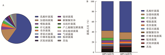
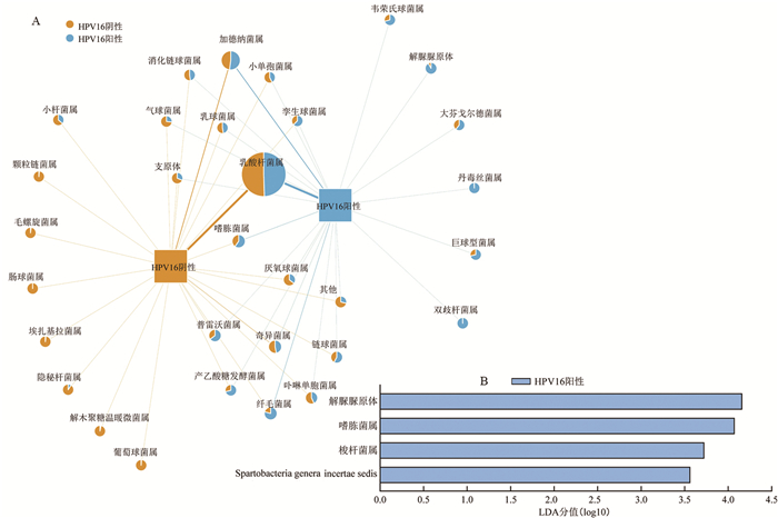
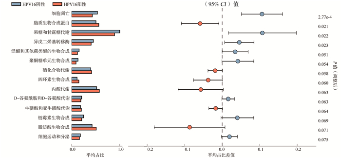
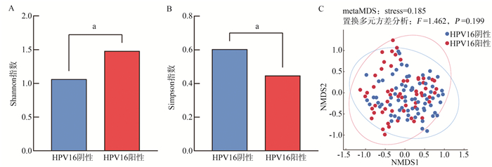
目的 探讨宫颈细胞学异常妇女不同HPV16感染状态下阴道菌群的特征,并对菌群功能进行预测。 方法 选取2018年1―6月在山西医科大学第二医院妇科门诊经液基薄层细胞学检查(liquid-based thinprep cytologic test, TCT)宫颈细胞学异常的132例妇女为研究对象,利用导流杂交法进行HPV感染及分型检测,采集阴道分泌物进行16S rDNA测序,利用生物信息学方法描述阴道菌群特征,并结合Kyoto Encyclopedia of Genes and Genomes(KEGG)数据库对阴道菌群功能进行分析。 结果 HPV16阳性妇女(59例)Simpson指数低于阴性妇女(73例)(P=0.047),而Shannon指数高于阴性妇女(P=0.036),基于Bray-Curtis距离的非度量多维尺度分析(non-metric multidimensional scaling, NMDS)显示两组Beta多样性差异无统计学意义(P=0.199)。宫颈细胞学异常妇女以及不同HPV16感染状态下均以乳酸杆菌为优势菌,但在HPV16阳性组乳酸杆菌占比降低(57.50%vs.60.69%),加德纳菌属(14.29%vs.13.66%)、嗜胨菌属(4.28%vs.2.99%)等厌氧菌属的构成比增加。在HPV16阴性组中乳酸杆菌属、肠球菌属、葡萄球菌属等丰度均高于阳性组,而HPV16阳性组中加德纳菌属、解脲脲原体、嗜胨菌属、普雷沃菌属等厌氧菌的丰度则均高于阴性组。Linear discriminant analysis Effect Size(LEfSe)软件分析显示解脲脲原体、嗜胨菌属为HPV16阳性组的特征菌。功能分析显示阴道菌群的功能主要集中在代谢层面,在HPV16阳性组以破坏阴道微环境和促进恶性肿瘤发生为主,HPV16阴性组以抑癌作用和糖脂代谢为主。 结论 阴道菌群多样性增加、比例结构改变与HPV16感染密切相关,解脲脲原体、嗜胨菌属可作为HPV16感染的潜在生物标志,HPV16感染状态下阴道菌群可能具有引发阴道微环境紊乱和促癌的功能。 -
关键词:
- 宫颈细胞学异常 /
- 人乳头瘤病毒16型 /
- 阴道菌群 /
- 16S rDNA测序 /
- 功能预测
Abstract:Objective To explore the characteristics of vaginal microbiota in women with abnormal cervical cytology under different HPV16 infection status, and to predict the function of the vaginal microbiota. Methods A total of 132 women with abnormal cervical cytology by liquid-based thinprep cytologic test (TCT), who came from the gynecological clinic of Second Hospital of Shanxi Medical University during January to June 2018, were enrolled in this study. Flow-through Hybridization technology was used to determine HPV infection typing. The vaginal microbiota was detected by 16S rDNA sequencing. The characteristic of vaginal microbiota were described by bio informatics, and Kyoto Encyclopedia of Genes and Genomes (KEGG) database was used to analyze the function of the vaginal microbiota. Results The Simpson index in the HPV16 positive group (n=59) was lower than that in the negative(n=73) group (P=0.047), while the Shannon index was higher in the positive group (P=0.036). Non-metric multidimensional scaling (NMDS) based on Bray-Curtis dissimilarities showed that there was no significance in Beta diversity between the two groups (P=0.199). Lactobacillus was the dominant bacteria in women with abnormal cervical cytology and different HPV16 infection status, but the proportion of Lactobacillus decreased (57.50%vs.60.69%) and anaerobic bacteria such as Gardnerella (14.29%vs.13.66%), and Peptoniphilus (4.28%vs.2.99%) increased in the HPV16 positive group. The abundance of Lactobacillus, Enterococcus, Staphylococcus and so on in the negative group was higher than that in the positive group, while the abundance of anaerobic bacteria such as Gardnerella, Ureaplasma, Peptoniphilus, Prevotella and so on in the HPV16 positive group was higher than that in the negative group. Ureaplasma and Peptoniphilus were the characteristic bacteria in HPV16 positive women based on Linear discriminant analysis Effect Size (LEfSe) analysis. The functional analysis showed that the function of vaginal microbiota was mainly concentrated on metabolism. The vaginal microbiota function in the HPV16 positive group was mainly to destroy the vaginal microenvironment and promote cell carcinogenesis, while the negative group was mainly to metabolism and inhibit cancer. Conclusions Increased diversity and the composition variation of vaginal microbiota are closely related to HPV16 infection. Ureaplasma and Peptoniphilus might be considered as potential biomarkers for HPV16 infection. Vaginal microbiota under HPV16 infection might induce vaginal microenvironment disorder and promote cancer. -

图 1 HPV16不同感染状态下阴道菌群多样性比较图
注:A:HPV16阳性与阴性组Alpha多样性Shannon指数比较;B:HPV16阳性与阴性组Alpha多样性Simpson指数比较;C:HPV16阳性与阴性组Beta多样性比较; aP < 0.05。
Figure 1. Comparison of diversity of vaginal microbiota among different HPV16 infection status

图 2 宫颈细胞学异常妇女及不同HPV16感染状态下阴道菌群的分布特征
注:A:宫颈细胞学异常妇女阴道菌群总体分布;B:不同HPV16感染状态阴道菌群结构。
Figure 2. Distribution characteristics of vaginal microbiota in women with cervical cytology abnormalities and different HPV16 infection status

图 3 HPV16不同感染状态下阴道菌群差异分析
注:A:不同HPV16感染状态阴道菌群Network图,饼图面积代表该菌丰度大小,颜色反映不同HPV16感染状态下该菌的丰度大小;B:不同HPV16感染状态LEfSe分析柱状图。
Figure 3. Analysis on the difference of vaginal microbiota under different HPV16 infection status
-
[1] Sung H, Ferlay J, Siegel RL, et al. GLOBAL cancer statistics 2020: globocan estimates of incidence and mortality worldwide for 36 cancers in 185 countries[J]. CA Cancer J Clin, 2021, 71(3): 209-249. DOI: 10.3322/caac.21660. [2] Stratton P, Battiwalla M, Tian X, et al. Immune response following quadrivalent human papillomavirus vaccination in women after hematopoietic allogeneic stem cell transplant: a nonrandomized clinical trial[J]. JAMA Oncol, 2020, 6(5): 696-705. DOI: 10.1001/jamaoncol.2019.6722. [3] Wong AK, Chan RC, Nichols WS, et al. Human papillomavirus (HPV) in atypical squamous cervical cytology: the Invader HPV test as a new screening assay[J]. J Clin Microbiol, 2008, 46(3): 869-875. DOI: 10.1128/JCM.01424-07. [4] Castle PE, Solomon D, Schiffman M, et al. Human papillomavirus type 16 infections and 2-year absolute risk of cervical precancer in women with equivocal or mild cytologic abnormalities[J]. J Natl Cancer Inst, 2005, 97(14): 1066-1071. DOI: 10.1093/jnci/dji186. [5] Kaambo E, Africa C, Chambuso R, et al. Vaginal microbiomes associated with aerobic vaginitis and bacterial vaginosis[J]. Front Public Health, 2018, 6: 78. DOI: 10.3389/fpubh.2018.00078. [6] Norenhag J, Du J, Olovsson M, et al. The vaginal microbiota, human papillomavirus and cervical dysplasia: a systematic review and network meta-analysis[J]. BJOG, 2020, 127(2): 171-180. DOI: 10.1111/1471-0528.15854. [7] Cohen PA, Jhingran A, Oaknin A, et al. Cervical cancer[J]. Lancet, 2019, 393(10167): 169-182. DOI: 10.1016/S0140-6736(18)32470-X. [8] 李俐, 丁玲, 吕元婧, 等. 阴道微环境改变与HPV16感染在宫颈上皮内瘤变中的作用及其交互效应[J]. 中华流行病学杂志, 2018, 39(11): 1486-1490. DOI: 10.3760/cma.j.issn.0254-6450.2018.11.013.Li L, Ding L, Lyu YJ, et al. Interaction between vaginal micro-environment alterations and HPV16 infection in cervical intraepithelial neoplasia[J]. Chin J Epidemiol, 2018, 39(11): 1486-1490. DOI: 10.3760/cma.j.issn.0254-6450.2018.11.013. [9] Di Paola M, Sani C, Clemente AM, et al. Characterization of cervico-vaginal microbiota in women developing persistent high-risk Human Papillomavirus infection[J]. Sci Rep, 2017, 7(1): 10200. DOI: 10.1038/s41598-017-09842-6. [10] Mitra A, MacIntyre DA, Ntritsos G, et al. The vaginal microbiota associates with the regression of untreated cervical intraepithelial neoplasia 2 lesions[J]. Nat Commun, 2020, 11(1): 1999. DOI: 10.1038/s41467-020-15856-y. [11] Wei ZT, Chen HL, Wang CF, et al. Depiction of vaginal microbiota in women with high-risk human papillomavirus infection[J]. Front Public Health, 2020, 8: 587298. DOI: 10.3389/fpubh.2020.587298. [12] Brotman RM, Shardell MD, Gajer P, et al. Interplay between the temporal dynamics of the vaginal microbiota and human papillomavirus detection[J]. J Infect Dis, 2014, 210(11): 1723-1733. DOI: 10.1093/infdis/jiu330. [13] Santiago GL, Deschaght P, El Aila N, et al. Gardnerella vaginalis comprises three distinct genotypes of which only two produce sialidase[J]. Am J Obstet Gynecol, 2011, 204(5): 450. e1-e450, e4507. DOI: 10.1016/j.ajog.2010.12.061. [14] Chen Y, Qiu X, Wang W, et al. Human papillomavirus infection and cervical intraepithelial neoplasia progression are associated with increased vaginal microbiome diversity in a Chinese cohort[J]. BMC Infect Dis, 2020, 20(1): 629. DOI: 10.1186/s12879-020-05324-9. [15] Robinson JW, Dando SJ, Nitsos I, et al. Ureaplasma parvum serovar 3 multiple banded antigen size variation after chronic intra-amniotic infection/colonization[J]. PLoS One, 2013, 8(4): e62746. DOI: 10.1371/journal.pone.0062746. [16] Moodley J, Constant D, Mwaka AD, et al. Mapping awareness of breast and cervical cancer risk factors, symptoms and lay beliefs in Uganda and South Africa[J]. PLoS One, 2020, 15(10): e0240788. DOI: 10.1371/journal.pone.0240788. [17] Farr Zuend C, Noël-Romas L, Hoger S, et al. Influence of dapivirine vaginal ring use on cervicovaginal immunity and functional microbiome in adolescent girls[J]. AIDS, 2021, 35(3): 369-380. DOI: 10.1097/QAD.0000000000002751. [18] Gonzalez PS, O'Prey J, Cardaci S, et al. Mannose impairs tumour growth and enhances chemotherapy[J]. Nature, 2018, 563(7733): 719-723. DOI: 10.1038/s41586-018-0729-3. [19] Loftus RM, Assmann N, Kedia-Mehta N, et al. Amino acid-dependent cMyc expression is essential for NK cell metabolic and functional responses in mice[J]. Nat Commun, 2018, 9(1): 2341. DOI: 10.1038/s41467-018-04719-2. -

 下载:
下载:
 点击查看大图
计量
- 文章访问数: 252
- HTML全文浏览量: 163
- PDF下载量: 31
- 被引次数: 0